On April 28, a research paper titled Structural Basis of GABAB Receptor-Gi Protein Coupling was published by Prof. Liu Jianfeng's research team under the Key Laboratory of Molecular Biophysics of MOE, College of Life Science and Technology, Huazhong University of Science and Technology, jointly with Prof. Zhang Yan's research team under the School of Medicine, Zhejiang University. The paper presented a breakthrough in identifying the structure of the class-C heterodimer GABAB receptor complexed with the G protein by high-resolution cryo-electron microscopy. The study, for the first time in the world, reveals a new mode of the dimer GPCR coupled with the G protein, which is of important guiding significance for the development of new GPCR allosteric drugs with less toxic and side effects.
As the largest family of membrane receptor proteins in human body, G protein-coupled receptors (GPCRs) play an important role in cell signal transmission and are closely related to human diseases. GPCRs are important drug targets, and more than 40% of the marketed drugs are developed with GPCRs as the target. GPCRs can be divided into four classes, i.e., A, B, C, and F, according to their similarity. GPCRs as nucleotide exchange factors can promote G protein to transform from GDP form to GTP form by coupling with the G protein, and thus activate downstream signaling. Previous studies have shown that classes A, B, and F monomer GPCRs move relative to transmembrane helix (TM) 3 through TM6, and make C-terminals of Gα subunits interact with each other in cavities on the inside of the receptor cell. More and more evidences show that different classes of GPCRs can form dimers or polymers, and class C GPCRs have been proved to function only through dimers formed by two identical or similar subunits. One GPCR dimer can only activate one G protein at a time, but the molecular mechanism of this asymmetric activation is not clear. Elucidation of this mechanism will enrich the new molecular pharmacology theory based on the GPCR dimer, and help to establish a new high-throughput drug screening method based on receptor dimer.
The GABAB receptor falls under the class C GPCRs, which is a heterodimer composed of subunits GB1 and GB2, and it is an important drug target for psychiatric diseases. This study reveals that, unlike other GPCRs, there is no TM6 expansion in the transmembrane regions of the two subunits under the activation of GABAB receptor, and only the GB2 subunit makes slight TM3 and TM5 movement, leading to the change of the intracellular loop (ICL) and the forming of a shallow groove by the TM3 and three ICLs to bind Gi protein. In addition, experiments show that the binding of the G protein to a GABAB receptor is a very flexible weak interaction. As shown in the figure, the binding of a GABAB receptor and the G protein is completely different from other types of GPCRs, indicating the uniqueness of asymmetric activation of the G protein by a GPCR dimer. The unique binding pattern between the GABAB receptor and the G protein has been further verified by many functional experiments, and this binding pattern may be conserved in other class-C GPCR dimers. The activated G protein retained all the expected conformational changes of an activated G protein: The α5-helix moves outward, and relatively, the helix domain moves, thus the nucleotide binding site is opened. The C-terminal of the α5-helix of the Gi protein is inserted between the long ICl2 and the short ICL3 of the GB2 subunit. This binding mode provides an explanation for the Gi/o selectivity of the GABAB receptor, and provides a structural basis for exploring the mechanism of the functional diversity of GPCRs. It is proposed for the first time that the steric hindrance between G proteins is the reason why a GPCR dimer only binds to one G protein, which solves a big problem in this field.
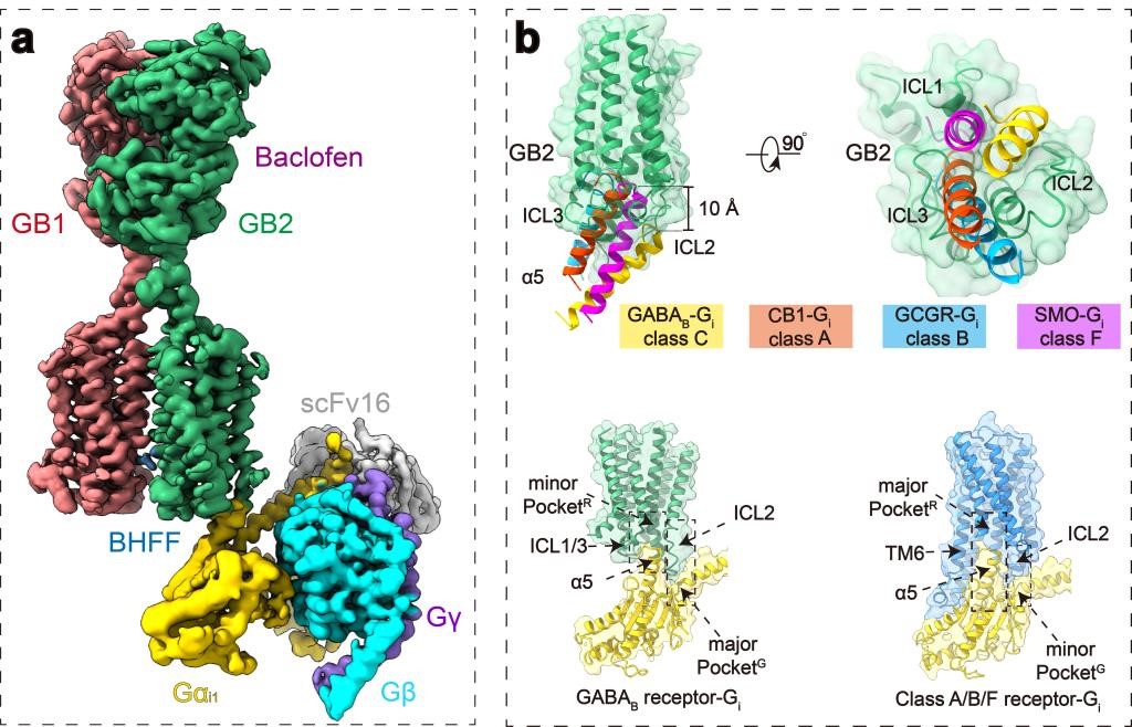
Fig. (a) shows the structure of a GABAB receptor complexed with a G protein under cryo-electron microscopy (Fig. a), and Fig. (b) shows the comparison between the GABAB receptor binding to a G protein and classes-A/B/F GPCRs binding to a G protein.
For the first time in the world, Prof. Liu Jianfeng and Prof. Zhang Yan's research teams analyzed at high resolution the structure of the GABAB receptor heterodimer in different states under the cryo-electron microscope in 2020, showing that the binding with the GABAB receptor agonist caused the closure of the VFT of GB1, which then induced TM interface rearrangement between the two subunits, and make TM transform from the inactive TM3-TM5/TM3-TM5 interface to the active TM6/TM6 interface (Cell Research, 2020). On the basis of previous studies, this work further illustrates the new mechanism of the receptor coupling with the G protein after the "bending" of GB2 by the receptor through the interaction between the extracellular regions of GB1 and GB2 and consequently the conformational change of GB2 transmembrane region, where the TM6/TM6 interaction between GB2 subunits stabilizes the "bent" state of the GB2 subunits, and then stabilizes the receptor activation. This work is the continuation of previous research, and describes the conformational changes of the GABAB receptor in different domains from the inactive state to the activated and coupled-with-G-protein state, which is of guiding significance to studies on activation of other GPCRs of the class C.
Liu Jianfeng's research team has been engaged in the GPCR molecular pharmacology studies for a long time, and has conducted systematic and in-depth research on the structure, function, and signal pathway of GPCR homodimers and heterodimers. The team has published more than 70 research papers on Cell, NAT Chem Biol., NAT Commun., SCI Adv., PNAS and other journals. In this work, class-C GPCR dimers previously discovered by Prof. Liu Jianfeng's research team are also further verified, such as activation modes of metabotropic glutamate receptors (NAT Chem Biol., 2015; PNAS., 2011), GABAB receptors (Cell Research, 2020; Nat Commun.,2019; EMBO J., 2008), and calcium sensitive receptor (PNAS., 2020). This study is another important achievement of joint cooperation between Zhejiang University and Huazhong University of Science and Technology since the establishment of the ZJU-HUST Joint Laboratory of Cellular Signaling. Shen Cangsong, a doctoral student at the School of Life Science and Technology of the HUST; Mao Chunyou, a postdoctor at the School of Medicine of the ZJU; Xu Chanjuan, a teacher at the School of Life Science and Technology of the HUST; and Jin Nan, a doctoral student at the School of Life Science and Technology, are the first authors of this paper. Prof. Liu Jianfeng at the School of Life Science and Technology, Prof. Jean-Philippe Pin at the French National Centre for Scientific Research, and Prof. Zhang Yan at the School of Medicine of Zhejiang University are corresponding co-authors. The project is supported by the national Key R&D Program of the Ministry of Science and Technology of China and the National Natural Science Foundation of China.
Link to the paper: Https://dx.doi.org/10.1038/s41586-021-03507-1

 Chinese