On February 3, 2023, a protocol paper titled “A practical guide to deep-learning light-field microscopy for 3D imaging of biological dynamics” was published in STAR Protocols by Prof. Fei Peng’s team at School of Optical and Electronic Information, Huazhong University of Science and Technology (HUST). This article presents a step-by-step protocol for the implementation of deep-learning-enhanced light-field microscopy enabling 3D imaging of instantaneous biological processes.
Current 3D imaging techniques face a significant challenge in extracting 3D information from millisecond transient cellular processes across long timescales, such as heartbeat, blood flow, neural activity, etc. Although several state-of-the-art microscopes, including epifluorescence and plane illumination methods, can achieve high-resolution 3D imaging of live samples, there is a trade-off between spatial resolution and volumetric imaging speed due to the axial scanning of multiple planes required to create a 3D volume.
To overcome these limitations, Fei Peng’s team provides a protocol for self-developed deep-learning-enhanced light-field microscopy capable of single-cell resolution 3D imaging at hundred-hertz volumetric imaging speed. This technology is a robust and versatile imaging tool for studying dynamics on fast timescales, potentially benefiting various applications such as behaviorally relevant neuronal activity studies and dysfunctions of the heart and blood transport system in model organisms. In addition to the superior performance, it should be also noted that VCD-LFM procedure is based on a relatively simple optical system and a spreadable computation framework, making it readily accessible to regular biological laboratories.
This protocol first provides the instructions to build a light-field microscope capable of capturing optically-encoded dynamic signals. Furthermore, the article details the data-processing and model training based on the user-friendly software package, which enables instant 3D image reconstruction from single 2D light-field snapshot. Finally, the protocol describes VCD-LFM imaging of several model organisms and demonstrates image-based quantitative studies on neural activities and cardio-hemodynamics (Fig. 1).

Fig. 1 Protocols for the implementation of deep-learning-enhanced light-field microscopy
For wider application, the design of light field imaging system provided in this paper is simple and robust (Fig. 2). The light-field microscope is based on a simple retrofit of a commercial wide-field microscope, which is convenient to fix and manipulate samples. All of the optical and optical mechanical components are off-the-shelf and constructed by cage system.
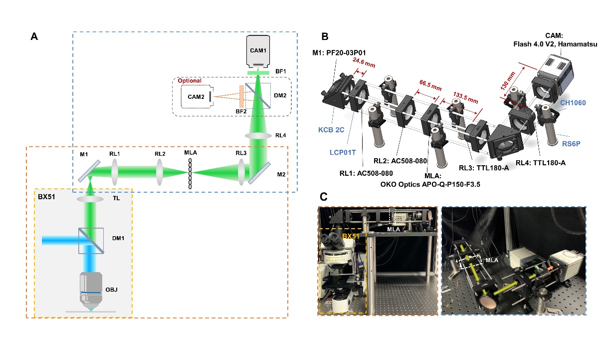
Fig. 2 Principle and prototype of light-field microscope
Moreover, the article describes the pipeline of data pre-processing and network training. To generate training data without a complex system to in situ acquire high-resolution label data and light field data, a special data preprocessing pipeline is illustrated to synthesize all training data only from the high-resolution label data. In addition, to better adapt the imaging requirements of users, two variations of the network structures are provided. A network with higher fitting ability handles structural signals (e.g., beads, red blood cells, myocyte nuclei and myocardium) and a lightweight network deals with functional imaging (e.g., calcium signals of C. elegans neurons) using a smaller model size while achieving higher inference speed.
Last but not least, the protocol describes the imaging protocol of several model organisms and demonstrates image-based quantitative analysis on neural activities and cardiac hemodynamics (Fig. 3).
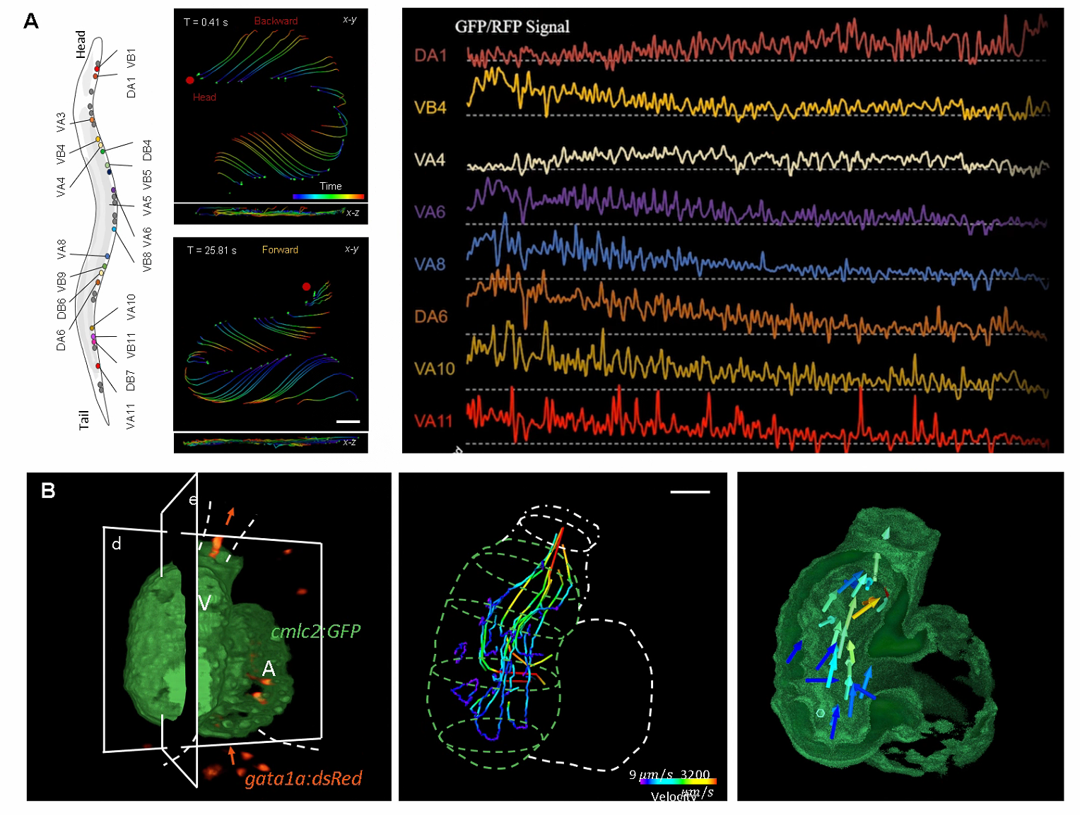
Fig. 3 Image-based quantitative analysis on neural activities and cardiac hemodynamics
Professor Fei Peng is the corresponding author of this paper. Zhu Lanxin is the co-corresponding author and first author of this article. Yi Chengqiang is the co-first author of the article.
Paper link: https://doi.org/10.1016/ j.xpro.2023.102078
Source: School of Optical and Electronic Information
Edited by: Peng Yumeng

 Chinese